Principles of interpretation
Border Effacement (silhouette sign)
- Border effacement occurs when two structures of the same radiopacity are in contact, leading to the inability to distinguish their margin.
- Contrarily, if these two structures are separated by a substance of a differing radiopacity, their borders can be distinguished radiographically.
To illustrate this principle we have taken a radiograph of two containers. These containers were positioned one on top of the other. The container on the bottom was filled with grapes and the container on top was filled with water
Photo of the containers
Radiograph of the containers
Water
Grapes & gas (air)
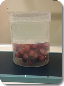
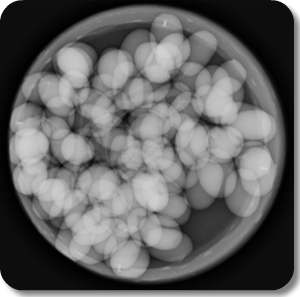
Note that the contours of the grapes can be identified. Even though there was a layer of water on top of the grapes, these were surrounded by gas and therefore their contours are visible
Animation showing the profile generated after the exposure of the grapes surrounded by gas. The grapes can be identified because more x-rays are being absorbed as they travel through both water and grapes compared to those being absorbed after traveling through only water
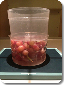
We have now decided to put both grapes and water in the same container (see photo) and take another radiograph. Assuming both have the same atomic number, what do you expect would happen with the radiographic appearance of the grapes?
Gas
Grapes & water
Click here to see the radiograph
Click here to see an animation of the exposure profile generated when grapes are surrounded by water
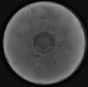
Because grapes and fluid have a similar atomic number, and in this case are also in contact, their contour has disappeared
In this case, regardless if x-rays are traveling through only fluid or fluid and grapes; a similar amount of x-rays are being absorbed, producing a flat exposure profile and an homogeneously gray radiograph
Radiographic geometry
Radiographic imaging are two-dimensional although the patient is three-dimensional
This means that the radiographic appearance of structures and/or lesions will depend on their orientation with respect to the primary x-ray beam and receiver
Consequences of these are magnification, distortion and supperimposition
Magnification
Magnification refers to the enlargement of a structure in the image relative to its actual size. Magnification depends mainly on the distance between the object and the receiver; as this distance increases, magnification increases
Animation showing the effect of changing the distance between the object and the source of x-ray on the size and sharpness of the generated image
Distortion
Distortion is unequal magnification that occurs when the object and receiver planes are not parallel.
Distortion leads to misrepresentation of the true shape or position of an object.
Some distortion occurs in every radiograph because there are always some parts of the patient that are not parallel to the plane of the receiver
Animation showing the effect of changing the orientation of an object in relation to the x-rays on the shape of the generated image
Animation showing the effect of changing the position of an object in relation to the x-rays on the shape of the generated image
Dr Mariano Makara
Dip. ECVDI